Saturday, March 22, 2014

Immature Granulocytes in PBFs

Immature Granulocytes in PBFs

1. Myeloblast


Background Information of Myeloblast

The myeloblast is the first recognizable unipotent stem cell (most immature), which will differentiate into one of the effectors of the Granulocyte series and this process is called granulopoiesis. The granulopoiesis consists of 5 stages, with the next differentiation sequence being pro-myelocyte. This cell have the ability to differentiate into one of the three different precursor cells, the neutrophilicbasophilic or eosinophilic myelocyteThe myeloblast is exclusively found in the bone marrow in normal circumstances and will not be found on PBFs. Presence of this cell type will indicate severe hematological disorders. 

Cellular Description


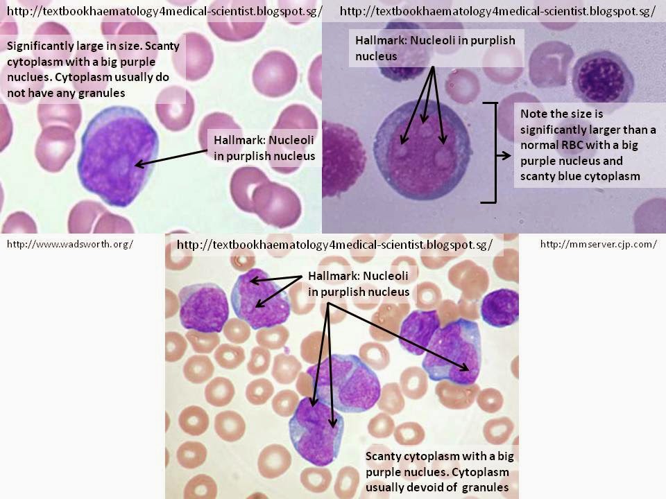
The hallmark of the myeloblast is the presence of 2-5 nucleoli that are brighter or lightly stain compared to their surrounding nucleus. The cell commonly appear as a significantly large round shape (maybe irregular at times) compared to the RBCs with relatively scanty blue cytoplasm and a large irregular purple nucleus. The cytoplasm is usually devoid of granules and the nucleus is composed of very fine non-aggregated chromatin.

2. Promyelocyte


Background Information of Promyelocyte

The Promyelocyte is the second differentiation sequence after differentiation from myeloblast in the granulopoiesis after differentiation.

Cellular Description

The hallmark of the Promyelocyte is the presence of coarse azurophilic (purplish) granules in the cytoplasm and may overlie the nucleus. The size of promyelocyte is usually larger than myeloblast with a reduced nuclear:cytoplasm ratio, meaning that the cytoplasm is more plentiful with a relatively smaller round/oval shaped nucleus compared to myeloblast. The cytoplasm staining varies between pinkish to bluish but the nucleus remain dark purple and the nucleus is usually skewed to a side of the cell (ie: not in the middle). Occasionally, nucleoli can still be present in the nucleus.  

3. Myelocyte


Background Information of Myelocyte

The Myelocyte is the third differentiation sequence after differentiation from Promyelocyte in the granulopoiesis after differentiation.

Cellular Description

The hallmark of the Myelocytes is the presence of fine lightly stained granules in the pinkish plentiful cytoplasm with a much relatively smaller round nucleus than both Promyelocyte and Myeloblast. The chromatin in the nucleus is stained more darkly in purple as it is more condensed and should not have any nucleoli. Furthermore, the nucleus is usually lying eccentrically. Myelocytes are significantly smaller than promyelocytes and have a 1:1 ratio of nucleus:cytoplasm.

4. Metamyelocyte


Background Information of Metamyelocyte

The Metamyelocyte is the fourth differentiation sequence after differentiation from Myelocyte in the granulopoiesis after differentiation. This is the last stage where these cells are considered immature.

Cellular Description

The hallmark of the Metamyelocyte is the presence of a large kidney-shaped or broad V shape nucleus in pinkish plentiful cytoplasm at the ratio of 1:1, cytoplasm:nucleus. There will not be any nucleoli in the nucleus nor there will be coarse granules in the cytoplasm. There will be fine lightly stained granules. It is the smallest in size in the immature granulocyte series when compared to myeloblast, promyelocyte and myelocyte, but almost similar in size with segmented/band neutrophils.

Discrepancy

Metamyelocytes are commonly mistaken as band neutrophils and vice-versa. It is a good practice to differentiate metamyelocytes from band neutrophils from the shape of nucleus. The nucleus of a band neutrophils has a much deeper band while a metamyelocyte has a shallow band.

Relative sizes of immature granulocytes to mature segmented neutrophils


Thursday, March 20, 2014

Sideroblastic anaemia

Sideroblastic anemia
Background Information


Sideroblastic anemia is a form anemia that is characterized by the presence of sideroblasts in the PBFs. Possible etiologies may either be a genetic disorder or indirectly as part of myelodysplastic syndrome, which can exacerbate into hematological malignancies (especially acute myelogenous leukemia). 
Patients with sideroblastic anemia do not have iron-deficiency, but the patients' bodies are unable to incorporate iron into the hemoglobin. Hence, RBCs may have granules of iron accumulate in the perinuclear mitochondria and nucleated RBCs may be present

The name Ring sideroblasts is suggestive that the iron granules are arranged in a ring form in the mitochondria around the nucleus. However this do not necessarily mean that a complete ring must be seen for a cell to be identified as a ring sideroblast.

According to WHO 2008 classification, three classification types of sideroblasts:
  1. Type 1 Sideroblasts: Fewer than 5 siderotic granules in the cytoplasm
  2. Type 2 Sideroblasts: 5 or more siderotic granules, but not in a perinuclear distribution
  3. Type 3 or Ring Sideroblasts: 5 or more granules in a perinuclear position, surrounding the nucleus or surrounding at least one third of the nuclear circumference.
Clinical Observations of PBFs
  • Dimorphic picture (presence of both normocytic and microcytic RBCs)
  • Hence MCV is commonly decreased but maybe normal
  • RDW is increased as there is marked poikilocytosis
  • Marked anisocytosis 
  • 20 - 30% hematocrit
  • Normal appearance and counts of both leukocytes and platelets
Further Tests  
  • Serum Iron: High
  • Increased ferritin Levels
  • Normal total iron-binding capacity (TIBC)
  • High transferrin saturation
  • Prussian Blue will reveal RBCs that contains blue-green granules (iron) in the cytoplasm. Prussian blue staining involves a non-enzymatic reaction of ferrous iron with ferrocyanide forming ferric-ferrocyanide, which is blue in color. A counter-stain may be used to provide better visualization.


Tuesday, March 18, 2014

Iron Deficiency Anemia

Iron Deficiency Anemia


Background Information


Iron-deficiency anemia is a common anemia characterized by the low RBC count or hemoglobin levels) caused by dietary deficiency and/or malabsorption of iron, and/or iron loss from internal bleeding which can originate from a range of sources such as the intestinal, uterine or urinary tract.
Iron deficiency causes approximately half of all anemia cases worldwide, and affects women more often than men.
This condition can surface if: 
  • The body does not produce adequate RBCs
  • Loss of RBCs more rapidly than they can be replaced probably due to bleeding
Often children in developing countries suffer from iron-deficiency anemia due to parasitic worms infection: hookworms, whipworms, and roundworms. These parasitic worms can inflict intestinal bleeding, which is not easily traceable in faeces, and is especially damaging to growing children. Malaria, hookworms and vitamin A deficiency are most common causes to this condition during pregnancy in most underdeveloped countries. For women after 50 years old, chronic gastrointestinal bleeding from non-parasitic causes, such as gastric ulcers, duodenal ulcers or gastrointestinal cancer are the most common cause of this condition.
Clinical Observations of PBFs
  • Low Hemoglobin and Hematocrit value
  • Increased RDW --> Anisocytosis
  • Low MCV (high number of abnormally small red blood cells) --> Microcytic RBCs
  • Low MCH and/or MCHC (Low hemoglobin --> Hypochromic)
  • Poikilocytosis: A plethora of variably-shaped RBCs - Target cells, hypochromic microcytic RBCs, Tear-Drop cells, elliptocytes and occasionally small numbers of nucleated RBCs.
  • platelet count is slightly above the high limit of normal (This is usually due to the proportionally reduced number of RBCs)


Megaloblastic Anemia

Megaloblastic Anemia


Background Information

Megaloblastic anemia is an anemia that results from inhibition of DNA synthesis during RBC production. this impaired DNA synthesis results in the cell cycle unable to progress from the G2 growth stage to the mitosis (M) stage. Hence the RBCs This leads continue to undergo cell growth without division, resulting in RBCs being larger than normal (macrocytosis) --> increased MCV. 

Megaloblastic anemia has a rather slow onset, especially when compared to that of other anemia. Hypovitaminosis, specifically a deficiency of vitamin B12 and/or folic acid leading to a malfunction in RBC DNA synthesis is often seen as the major etiology of megaloblastic anemia. However, Vitamin B12 deficiency alone will not cause the syndrome in the presence of sufficient folate, for the mechanism is loss of B12 dependent folate recycling, followed by folate-deficiency loss of nucleic acid synthesis, leading to defects in DNA synthesis. Fortunately, folic acid supplementation in the absence of vitamin B12 prevents this type of anemia (although other vitamin B12-specific pathologies continue). Additionally, copper deficiency resulting from zinc excess from unusually high oral consumption of zinc containing denture fixation creams has been found to be a cause.

Uncommonly, anti-metabolites such as some chemotherapeutic or antimicrobial agents (for example azathioprine or trimethoprim) that inhibit DNA production directly, can cause this condition too

Clinical Observations of PBFs
  • Reduced RBC count and hemoglobin levels
  • Increased MCV (>95 fl) due to macrocytes (larger sized biconcave round RBCs) and Ovalocytes (generally larger in size) seen and MCH (due to increase in hemoglobin concentration per cell)
  • Normal MCHC (32–36 g/dL)
  • The platelet count may be reduced.
  • Hyper-segmented Neutrophils may be observed. This is thought to be due to decreased production of "new" neutrophils, hence a compensatory prolonged lifespan for circulating neutrophils, which increase numbers of nuclear segments with age.
  • Increased RDW due to increased variation in RBC size (Anisocytosis)
  • Poikilocytosis (abnormally shaped RBCs): Macrocytes, Ovalocytes

Diagnostic Strategy
Once macrocytosis is identified, the history and physical examination help narrow the differential diagnosis.
*MMA = methylmalonic acid

Friday, March 14, 2014

Spherocytes

Spherocytes


Background Information of Spherocytes

Spherocytes are RBCs that have a molecular defect in 1 or more proteins in the RBC cytoskeleton. The cytoskeleton of RBCs are typically composed of Spectrin, Ankyrin, Band 3 or Protein 4.2. Due to their cytoskeleton defect, the RBC contracts to its most surface-tension efficient and least flexible configuration, which is the sphere-like shape (as the name implies). However this configuration is susceptible to high osmotic fragility and prone to physical degeneration.


Clinical Significance
Appearance of numerous spherocytes without other abnormal RBCs on a PBF is highly indicative of Hereditary spherocytosis.  If small numbers of spherocytes is seen in conjunction with other abnormal RBCs are seen in PBFs, the following clinical conditions may be suspected: 
  • Isoimmune and autoimmune hemolytic anemias 
  • Heinz body hemolytic anemia
  • Hereditary pyropoikilocytosis
  • Microangiopathic hemolytic anemia
  • Hypersplenism
  • Post-splenectomy
  • Myelofibrosis with myeloid metaplasia
  • Hemoglobinopathies
  • Malaria
  • Liver disease
  • recent transfusions
  • severe burns. 


Cellular Description

The hall mark of spherocytes is the presence of numerous small RBCs that appear synonymous to a sphere-like shape without a visible central pallor and is stained more densely than their normal counterparts. The denser staining is due to the normal concentration of hemoglobin being "concentrated" due to the shrinkage of the RBCs.

Thursday, March 13, 2014

Red Cell Indices

Red Cell Indices

Mean Corpuscular Volume (MCV)
It is the measure of the average RBC volume and RBC size. This red cell indices allow the classification of anaemia. The value of MCV increases proportionally with the size of the RBCs (ie bigger the cell size --> the higher the MCV value)

a)Normocytic anaemia
  • Usually a reduced production of normal RBC or increased destruction of RBC
  • Usually appear with reduced haematocrit and haemoglobin
a)Macrocytic anaemia
  • Megaloblastic anaemia
b)Microcytic anaemia
  • Iron Deficiency
  • Thalassemia
  • Sideroblastic Anaemia

Mean Corpuscular Hemoglobin (MCH)

Mean Corpuscular Hemoglobin (MCH) indicates the average mass of the hemoglobin per 
RBC in a sample of blood. The haemoglobin in RBCs is located primarily in the peripheral, 
leaving an area of pale staining area called the central pallor equal to approximately 30 to 
45% of the diameter of the RBCs. Hence if the central pallor is expanded (Hypochromatic), 
the concentration of the hemoglobin will then be reduced leading to a reduced MCH. On 
the contrary, if the size of the central pallor is reduced, the hemoglobin concentration will 
increase, so will the MCH.


Mean Corpuscular Hemoglobin Conentration (MCHC)


Mean Corpuscular Hemoglobin Conentration (MCHC) is the measure of the concentration 
of hemoglobin in a given volume of packed RBCs. This red cell indices allow a more 
in-depth classification of anemia. The value of MCHC is a correlation of concentration of 
hemoglobin and the size of the RBCs.


Sunday, March 9, 2014

Activated Partial Thromboplastin Time (APTT)

Activated Partial Thromboplastin Time (APTT)

Another blood clotting test, called partial thromboplastin time (PTT), might be used if you take another type of blood-thinning medicine called heparin. This test measures other clotting factors. Partial thromboplastin time and prothrombin time are often done at the same time to check for bleeding problems or the chance for too much bleeding in surgery.

Prothrombin Time (PT)

Prothrombin Time (PT)

BackGround Information
Prothrombin time (PT) is a hematological test that determines how long it takes for a patient's blood to clot. It measures the extrinsic pathway of coagulation. This test is traditionally used to check for bleeding problems. Additionally, PT can also be used to check the efficacy of medicines that aids in preventing blood clots.

Prothrombin Time International normalized ratio (PTINR)

PTINR (international normalized ratio) is commonly measured to standardize the results of prothrombin time tests, as different automated hematological coagulation anaylzers and reagents (tissue factor) will yield different results. Hence, this allows physicians to interpret results correctly even though patients' results may come from different laboratories and different test methods. Therefore this PTINR system permits the treatment with blood-thinning medicine (anticoagulant therapy) to be effective. 
* Each manufacturer assigns an ISI value (International Sensitivity Index) for every tissue factor they manufacture. The ISI value indicates how a particular batch of tissue factor compares to an international reference tissue factor. The ISI is usually between 1.0 and 2.0. The INR is the ratio of a patient's prothrombin time to a normal (control) sample, raised to the power of the ISI value for the analytical system used.
Clinical Significance
Prothrombin (factor II), is one of the crucial clotting factors made by the liver. Vitamin K is essential to produce prothrombin and other clotting factors. PT test is essential to determine the presence and functionality of five vital blood clotting factors (factors I, II, V, VII, and X). Abnormally prolonged PT is often a result of underlying liver disease or injury or by treatment with blood thinners.
Prothrombin Time can be prolonged due to circumstances like:

  • Blood-thinning medicine, such as warfarin.
  • Low levels of blood clotting factors (factors I, II, V, VII, and X).
  • A change in the activity/functionality of any of the clotting factors.
  • The absence of any of the clotting factors.
  • Other substances or inhibitors, that affect the clotting factors.
  • An increased usage of the clotting factors.
Procedures for Manual PT Test Method
  • Ensure appropriate volume of blood in sodium citrated tube (Blue-top)
  • Centrifuge sodium citrated tube at a speed suitable to obtain platelet poor plasma (< 10 X 103/μL)
  • Pre-warm sufficient amount of Dade Innovin (~1200ul to run in duplicates with Low and High QC) at 37°C for 15 minutes
  • Pipette 0.1ml (100ul) of citrated plasma into a test tube preheated to 37°C
  • incubate for 3 minutes at 37°C
  • Pipette 0.2ml (200ul) of prewarmed Dade Innovin into plasma
  • Immediately upon addition of Dade Innovin, start the stopwatch and determine the clotting time.
  • The result is the elapsed time, in seconds, from the addition of the Dade Innovin into the plasma until the appearance of a solid gel clot
Reference Range

Normal Range: 9.6 - 11.6 seconds